

Are you planning on performing IHC as part of your study? Is the data you obtain from IHC critical in your decision making process? As any scientist knows, it is very important to plan your experiment well to achieve high-quality, repeatable results.
IHC is a complicated process that requires planning ahead so that tissues are collected and fixed properly before you start. A good outsourcing partner can help you with your IHC and give you consistent repeatable results.
There are many steps and variables to be considered when planning your IHC:
Each step, from tissue collection through data analysis, will impact the outcome of your IHC study. Our very experienced and knowledgeable team of histologists, scientists, and pathologists can help you every step of the way.
Your first consideration is tissue collection and fixation
Depending on what marker you want to look at, your tissue fixation needs may vary. Most antibodies work well on formalin-fixed paraffin-embedded (FFPE) tissues, but some will only work on frozen tissues. After fixing your samples, they must be processed and embedded. Embedding in paraffin can be messy and requires familiarity with the procedure to avoid ruining crucial samples. If infiltration with paraffin is incomplete, you’re going to get damaged sections. Embedding in OCT, for frozen sections, can also be tricky. You need to ensure that there are no air bubbles in the media and that the OCT is being frozen properly. Once you have properly embedded samples, you will want an expert histologist to cut your sections. Getting tissue sections without wrinkles, tears, folds and other artifacts is very tricky, and requires an experienced hand. Spacing of tissue on a slide is also important, to allow for clean staining and to prevent cross-contamination.
Antibody selection is of utmost importance
Most proteins have multiple commercially available antibodies available on the market. How do you know which one to choose? This requires experience and some understanding of the principles of IHC. Some of the questions that we would ask are: Does the antibody react with the specific epitope in question? Has the antibody been tested in the species we’re researching? Is it a polyclonal or a monoclonal antibody? What is the host species of the antibody? What antigen retrieval method is best for my antibody and tissue? At what concentration will I need to use my antibody? What are the proper controls for my IHC? Will multiplexing be required?
These are just a few of the many questions and details that can go into optimization of an IHC protocol. That’s why you should trust repeatable protocols performed by experts using an automated staining system.
Consistent sample processing
What if you want to determine if two proteins co-localize within a cell? This requires careful optimization of two antibodies on the same tissue with similar conditions. If not done properly, this process can consume large quantities of antibodies and reagents. Consistency is key. Given the cost of antibodies and other reagents, you’re not going to want to be testing dilutions and developing protocols when you have important samples to process just waiting in the pipeline. Our team routinely performs dual immunofluorescent staining and has extensive experience.
Save money
Our protocol optimization system saves you money through efficient use of reagents. In addition, we can provide you with scanned images at reasonable costs, saving you from having to purchase your own imaging system.
Save time
You’ll save time by letting us process your samples, letting you get on with other aspects of your work. Because we have an experienced team, we can get your work done in timely and efficient manner, saving you both time and money.
Discuss your experimental design with experts
We have experienced pathologists and scientists available to discuss your study needs and endpoints to help you identify the best IHC markers to use to reach your goals. We have multiple protocols in place and are very experienced at developing new protocols to meet your needs.
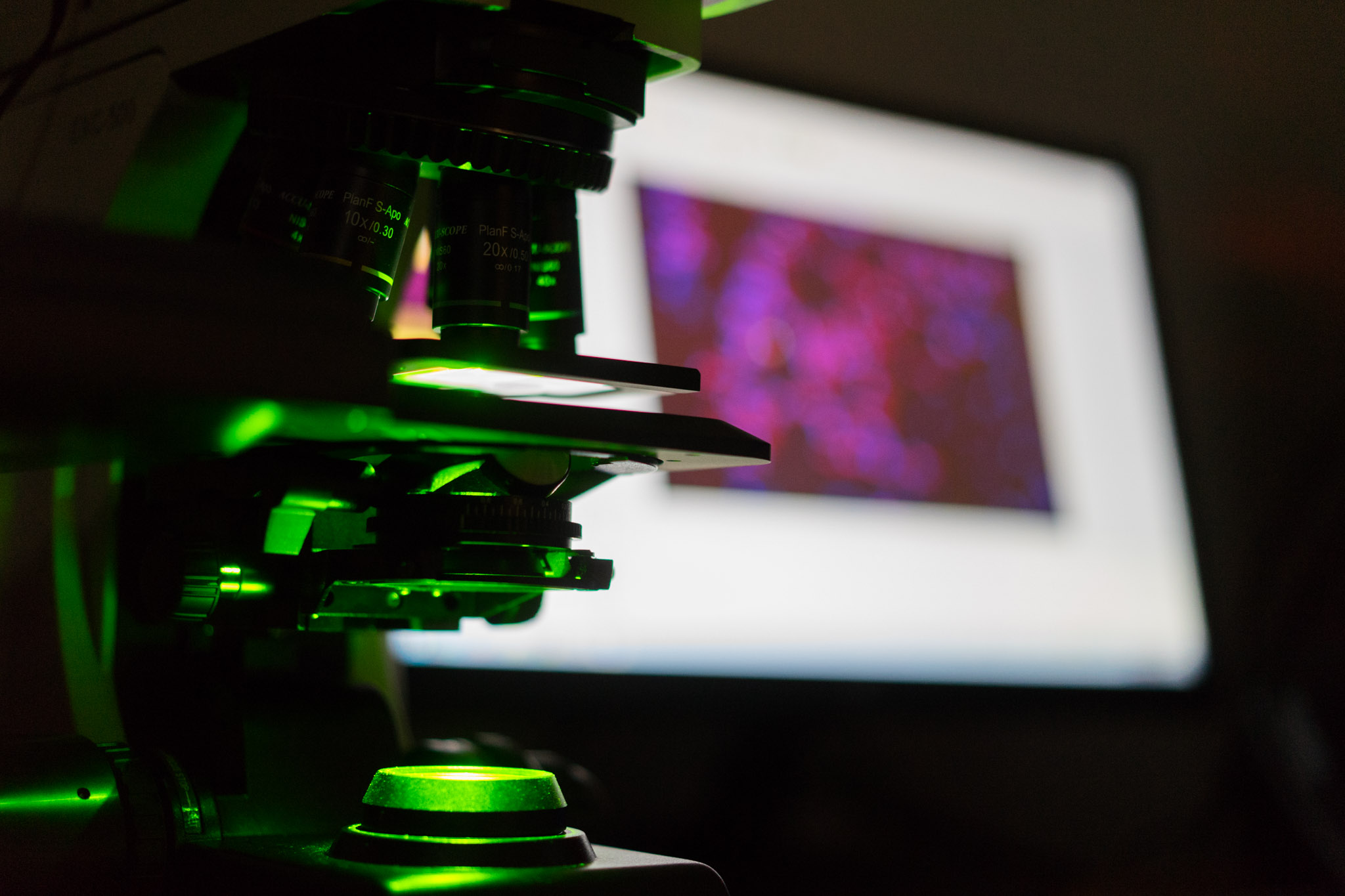
Imaging and Image Analysis
In addition to our IHC services, we provide slide scanning services and quantitative image analysis. A high-quality scanner can scan both chromogenic and fluorescent labeled slides. A good partner can provide you with whole slide images or can focus on a particular region of interest for imaging. An experienced team can provide you with exceptional quality images and data.
If you have any questions regarding IHC sample processing, please contact us at HSRL today. We’re happy to discuss the best options available for your specific experimental needs.